JOINTS 2023;
1: e594
DOI: 10.26355/joints_20235_594
When progress unmasks the dangerous violation of the wisdom of nature
Topic: Rehabilitation
Category: Editorial
Abstract
Some interventions, such as percutaneous fibrolysis, deep massages, performed on muscle tissues during the biological reparative period, can negatively interfere with the normal tissue reparative response, consequently altering the imaging control performed. Sometimes such therapies can cause iatrogenic damage to muscle tissues during its biological repair phase. Imaging examination, especially magnetic resonance imaging, can unmask these iatrogenic damages. The aim of this paper is to draw attention to the importance of careful evaluation concerning these therapies performed during the rehabilitation pathway, especially during the biological reparative period, since recent radiological techniques seem to show a higher frequency of iatrogenic lesions.In the use of imaging techniques, the last few years have been characterized by significant progress in imaging. Indeed, the two most used techniques in this area, namely magnetic resonance imaging (MRI) and ultrasonography (US), have made remarkable progress in the last decade1,2. In the study of muscle pathologies, modern MRI high-field strength magnets are able to support a higher spatial and contrast resolution more easily, being so able to more accurately characterize the muscle and tendon injuries, as well as the intramuscular connective tissue lesions3. Concerning the MRI, some techniques, such as diffusion-weighted imaging (DWI) and diffusion tensor imaging (DTI), seem to be particularly promising. DWI imaging allows the measurement of molecular diffusion and is particularly interesting in the study of muscle inflammation3. The DTI allows the tracing of muscle fibers, thus proving to be extremely suitable for the study of sub-structural muscle injuries4. However, for its greater spatial resolution and its greater contrast gradient, MRI represents the gold standard exam for the study of muscle-tendon pathologies5. Obviously, this enormous improvement in imaging techniques should not exempt the clinician from having a high clinical capacity and a deep knowledge of semeiotics. Indeed, only a deep theoretical and practical knowledge of the latter, together with an equally high interpretive ability of imaging, will allow the formulation of a precise diagnosis. Recent advances in imaging have also allowed a follow-up of the muscle injury healing process and have made it possible to highlight how the non-evidence-based use of some therapeutic interventions can negatively affect the normal biological repair processes of muscle tissue. For example, interventions such as percutaneous fibrolysis or deep massages, performed on tissues still in the process of biological reparative reworking, can negatively interfere with the normal tissue reparative response, consequently altering the imaging control performed. For this reason, the use of any non-evidence-based therapeutic intervention should be discouraged. Indeed, especially in the early stages, their use on muscle tissue still in the process of repair/regeneration can be extremely counterproductive.
The consequent imaging alterations are not only extremely misleading for the clinician, in the event that the latter is unaware of the treatments causing these radiological alterations (as often happens in football with many players embarking on non-evidence-based therapeutic paths), but they can also result in serious iatrogenic complications.
The assumption that the clinic is more important than has always been so cited that it has essentially become a medical dogma. However, given the important advances in imaging in recent years, to date this dogmatism, at least in some situations, may be questioned.
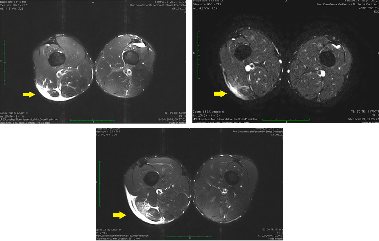
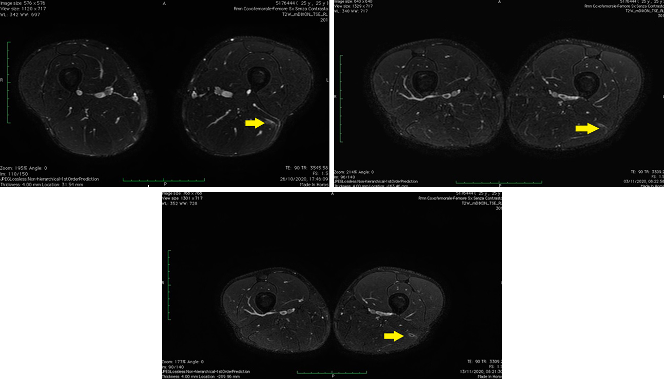
Two practical examples concerning previous discussions can be found in Figure 1 and Figure 2.
Figure 1. A, axial MRI STIR image showing a 2nd-degree injury of the right hamstring (arrow). B, axial STIR control image performed 12 days after the previous exam showing a clear reduction of the signal hyperintensity (arrow). C, axial control STIR image performed 14 days after the second examination, showing a dramatic increase in the signal hyperintensity zone in the area undergoing biological repairs (arrow). This radiological worsening is probably due to a deep massage performed in the area still being repaired. It should be noted that no re-injury event had occurred during the rehabilitation process.

Figure 2. A, Axial T2 MRI image showing a 1st-degree lesion of the left hamstring. B, axial T2 control image performed 8 days after the previous examination showing an almost total disappearance of the hyperintensity zone. C, T2 axial control image performed 10 days after the previous examination showing a reappearance of the signal hyperintensity in the regeneration zone (arrow). The radiological worsening is probably due to a percutaneous fibrolysis operation performed in the area still undergoing biological repair. As in the previous case no re-injury event had occurred.

Therefore, our message intends to draw attention to the fact that in sport medicine it is important to carefully evaluate all therapies before their administration to athletes, balancing the advantages and the (eventual) adverse events, costs and clinical gains. Indeed, recent radiological techniques seem to show a higher frequency of iatrogenic lesions after the administration of some non-evidence-based treatments.
Finally, we would like to add another reflection in our opinion very important from an ethical and deontological point of view.
Since these therapeutic treatments are performed in an attempt (often in vain) to accelerate the processes of tissue repair, we would like to quote in this regard a phrase by Hans Jonas, an important philosopher and authoritative exponent of the current of Gnosticism.
Jonas6, in his book entitled “Technique, medicine and ethics”, says: “Everyone must ask themselves whether it is good and wise, in consideration of the good of the individual and of the group, to bring confusion and ephemeral hedonism in the wisdom of nature which has established its times through a long evolution”. We would like to invite the reader to reflect on this sentence.
Conflict of Interest
The Authors declare no conflict of interest.
Funding
This research received no external funding
Availability of Data and Materials
The data presented in this work is available upon request from the corresponding author.
ORCID ID
Bisciotti Gian Nicola: 0000-0003-1346-320X
Volpi Piero. 0000-0001-7938-4964
Eirale Cristiano: 0000-0001-5928-7382
References
- Bordalo M, Arnaiz J, Yamashiro E, Al-Naimi MR. Imaging of Muscle Injuries: MR Imaging-Ultrasound Correlation. Magn Reson Imaging Clin N Am 2023; 31: 163-179.
- Heiss R, Janka R, Uder M, Hotfiel T, Gast L, Nagel AM, Roemer FW. Bildgebung von Muskelverletzungen im Sport [Imaging of muscle injuries in sports medicine]. Radiologie (Heidelb) 2023; 63: 249-258.
- Guermazi A, Roemer FW, Robinson P, Tol JL, Regatte RR, Crema MD. Imaging of Muscle Injuries in Sports Medicine: Sports Imaging Series. Radiology 2017; 282: 646-663.
- Martín-Noguerol T, Barousse R, Wessell DE, Rossi I, Luna A. A handbook for beginners in skeletal muscle diffusion tensor imaging: physical basis and technical adjustments. Eur Radiol 2022; 32: 7623-7631.
- Bisciotti GN, Volpi P, Amato M, Alberti G, Allegra F, Aprato A, Artina M, Auci A, Bait C, Bastieri GM, Balzarini L, Belli A, Bellini G, Bettinsoli P, Bisciotti A, Bisciotti A, Bona S, Brambilla L, Bresciani M, Buffoli M, Calanna F, Canata GL, Cardinali D, Carimati G, Cassaghi G, Cautero E, Cena E, Corradini B, Corsini A, D’Agostino C, De Donato M, Delle Rose G, Di Marzo F, Di Pietto F, Enrica D, Eirale C, Febbrari L, Ferrua P, Foglia A, Galbiati A, Gheza A, Giammattei C, Masia F, Melegati G, Moretti B, Moretti L, Niccolai R, Orgiani A, Orizio C, Pantalone A, Parra F, Patroni P, Pereira Ruiz MT, Perri M, Petrillo S, Pulici L, Quaglia A, Ricciotti L, Rosa F, Sasso N, Sprenger C, Tarantola C, Tenconi FG, Tosi F, Trainini M, Tucciarone A, Yekdah A, Vuckovic Z, Zini R, Chamari K. Italian consensus conference on guidelines for conservative treatment on lower limb muscle injuries in athlete. BMJ Open Sport Exerc Med 2018; 4: e000323.
- Jonas H. Tecnica, medicina ed etica. Einaudi (Eds) Torino, 1995; 59.
To cite this article
When progress unmasks the dangerous violation of the wisdom of nature
JOINTS 2023;
1: e594
DOI: 10.26355/joints_20235_594
Publication History
Submission date: 18 Apr 2023
Revised on: 30 Apr 2023
Accepted on: 03 May 2023
Published online: 26 May 2023